On Line Enhanced Content For Chapter 1 "History and Physical Examination" DERMATOLOGIC DESCRIPTIONS. This on line version includes some illustrative color images of the patterns described.
Atrophy: Thinning of the surface of the skin with associated loss of normal markings. Examples: Aging, striae associated with obesity, scleroderma
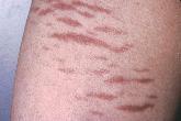
Striae
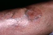
necrobiosis lipoidica diabeticorum (NLD)
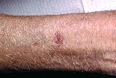
Solar Purpura
Bulla: A superficial, well-circumscribed, raised, fluid-filled lesion greater than 1 cm in diameter. Examples: Bullous pemphigoid, pemphigus, dermatitis herpetiformis
Burrow: A subcutaneous linear track made by a parasite. Example: Scabies Crust: A slightly raised lesion with irregular border and variable color resulting from dried blood, serum, or other exudate. Examples: Scab resulting from an abrasion, impetigo Ecchymosis: A flat, nonblanching, red-purple-blue lesion that results from extravasation of red blood cells into the skin. Differs from purpura in size; ecchymoses are large purpura lesions. Examples: Trauma, long-term steroid use Erosion: A depressed lesion resulting from loss of epidermis due to rupture of vesicles or bullae. Example: Rupture of herpes simplex blister Excoriation: A linear superficial lesion, which may be covered with dried blood. Early lesions with surrounding erythema. Often self-induced. Example: Scratching associated with pruritus from any cause Fissure: A deep linear lesion into the dermis. Example: Cracks seen in athlete’s foot Keloid: Irregular, raised lesion resulting from hypertrophied scar tissue. Examples: Often seen with burns; African Americans are more prone to keloid formation than are other people. Lichenification: A thickening of the skin with an increase in skin markings resulting from chronic irritation and rubbing. Example: Atopic dermatitis Macule: A circumscribed nonpalpable discoloration of the skin less than 1 cm in diameter. Examples: Freckles, rubella, petechiae Nodule: A solid, palpable, circumscribed lesion larger than a papule and smaller than a tumor. Examples: Erythema nodosum, gouty tophi Papule: A solid elevated lesion less than 1 cm in diameter. Examples: Acne, warts, insect bites Patch: A nonpalpable discoloration of the skin with an irregular border, greater than 1 cm in diameter. Example: Vitiligo Petechiae: Flat, pinhead-sized, nonblanching, red—purple lesions[CJA1] caused by hemorrhage into the skin. Example: Seen in DIC, ITP, SLE, meningococcemia (Neisseria meningitidis) Plaque: A solid, flat, elevated lesion greater than 1 cm in diameter. Examples: Psoriasis, discoid lupus erythematosus, actinic keratosis Purpura: A condition characterized by[CJA2] flat, nonblanching, red—purple lesions larger than petechiae caused by hemorrhage into the skin. Examples: Henoch–Schönlein purpura, TTP Pustule: A vesicle filled with purulent fluid. Examples: Acne, impetigo Scales: Partial separation of the superficial layer of skin. Examples: Psoriasis, dandruff Scar: Replacement of normal skin with fibrous tissue, often resulting from injury. Examples: Surgical scar, burn Telangiectasia: Dilatation of capillaries resulting in red, irregular, clustered lines that blanch. Examples: Seen in scleroderma, Osler–Weber–Rendu disease, cirrhosis Tumor: A solid, palpable, circumscribed lesion greater than 2 cm in diameter. Example: Lipoma Ulcer: A depressed lesion resulting from loss of epidermis and part of the dermis. Examples: Decubitus ulcers, primary lesion of syphilis, venous stasis ulcer Vesicle: A superficial, well-circumscribed, raised, fluid-filled lesion that is less than 1 cm in diameter. Examples: Herpes simplex, varicella (chickenpox) Wheal: Slightly raised, red, irregular, transient lesions secondary to edema of the skin. Examples: Urticaria (hives), allergic reaction to injections or insect bites